|
|
|
Health Matters Related to Periodontal Diseasebleeding gums contagious diabetes halitosis (breath) heart disease heredity info pages oral cancer cancer treatment periodontal disease pregnancy self test smoking- tobacco women Prevention Treatment Our Office Articles of interest in Periodontics
|
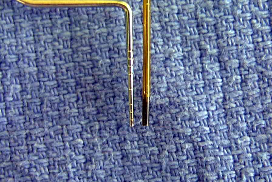
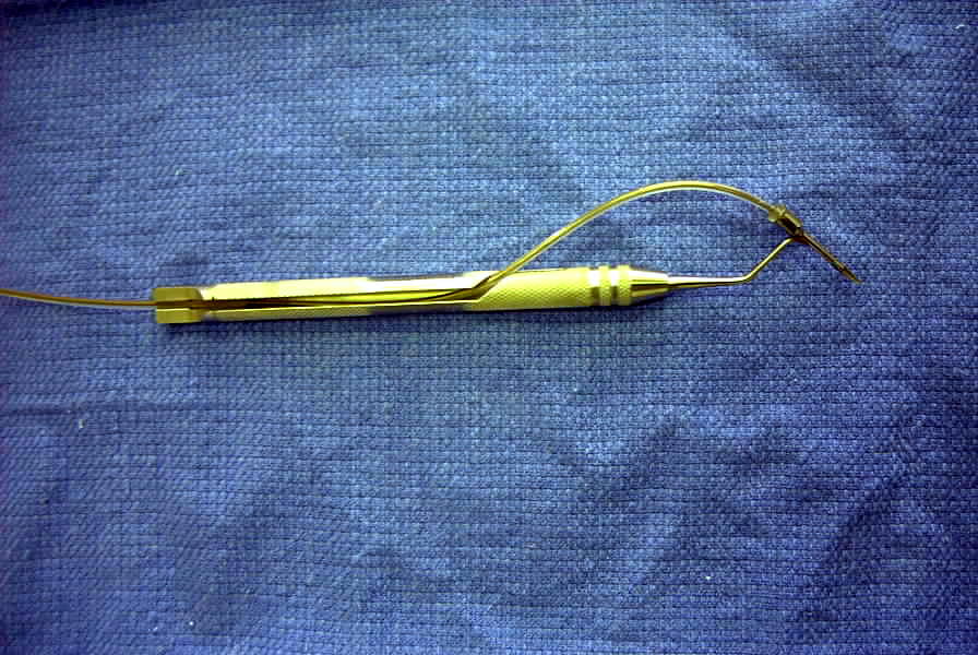
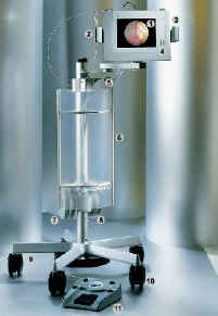
Before viewing any actual images from The Perioscope, it is important to understand something about the unit. The image above shows (from the floor up) a foot control which regulates irrigation flow and light intensity. The unit rests on a very sturdy wheelbase with lockable casters. A 3 inch diameter mounting post supports the main unit which houses the LCD monitor, the signal processor, the irrigation unit and the light source. The endoscope screws into the top of the unit. It is a bundle that contains fiber optic elements that transmit the video signal back to the processor as well at light from the unit to the object being viewed. The image below shows the size of the bundle compared to a periodontal probe.
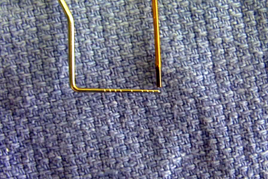
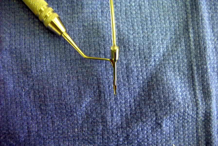
The bundle is inserted into a sterile sheath which insures asepsis. The sheath is biluminal and provides for irrigant flow to the tip without allowing the bundle to become moist. The irrigation system is crucial for viewing. Active hemorrhage is continually washed from the viewing field. The sheathed fiber optic bundle is then positioned into a hand instrument for intraoral use (see image below).
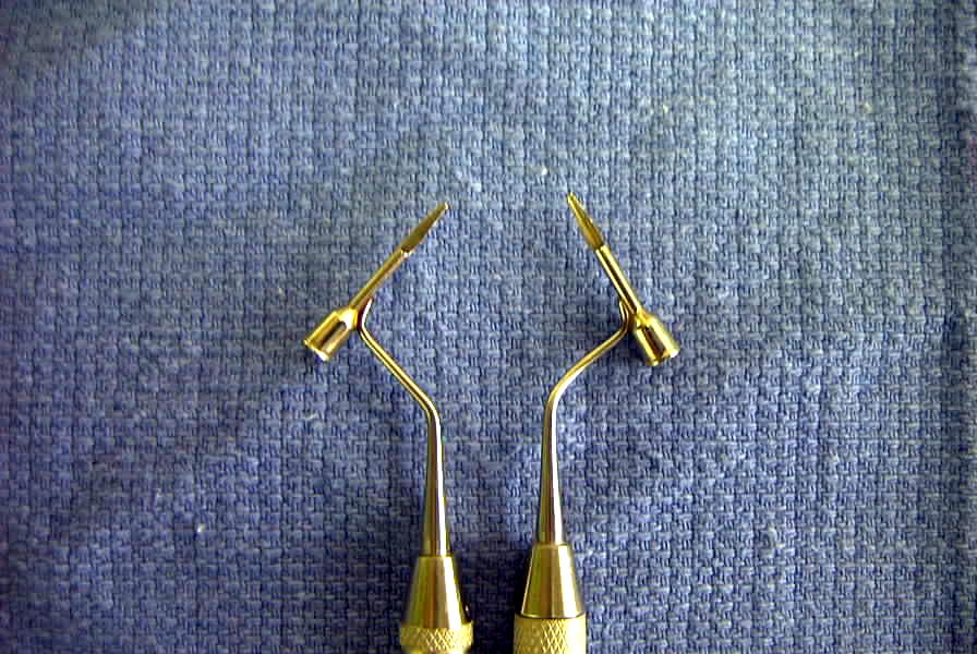
There are a variety of holders. The image below shows a right and a left holder. There are others including a straight and variants of the right and left with curves built in to improve mesial or distal viewing.
The fully assembled holder, sheath and fiber optic bundle are shown below.
Dental Endoscopy C.E. Review for Dental Professionals
As always, Aymee and I welcome your referrals and would be happy to discuss any case where you think endoscopy may be beneficial for your patient. Your confidence is appreciated!
|

 The Perioscopic Primer